


Unraveling the Myth: The Truth About the Kraken
Is the Kraken real yes or no? For centuries, sailors and seafaring enthusiasts have been captivated by tales of the legendary sea monster known as the …
Read Article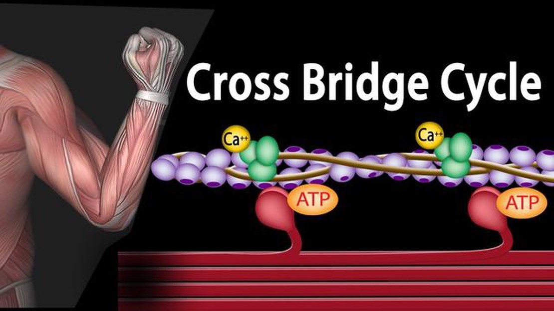
Muscle contraction is a complex physiological process that allows our bodies to move and perform various physical tasks. It involves a series of sequential steps that involve the interaction of different proteins and ions within the muscle fibers. Understanding the process of muscle contraction is crucial in fields such as sports science, rehabilitation, and even gaming, where precise muscular movements can make a difference in gameplay.
At the cellular level, muscle contraction starts with an electrical signal from the nervous system. When the brain sends a signal to initiate movement, an action potential is generated and travels down the motor neuron to reach the muscle fibers. This action potential then causes the release of calcium ions from the sarcoplasmic reticulum, a network of tubules within the muscle cell.
Once the calcium ions are released into the muscle cell, they bind to a protein called troponin, which is located on the actin filaments. This binding causes a change in the shape of the troponin-tropomyosin complex, exposing the myosin-binding sites on the actin filaments. The myosin heads, which are part of the thick filaments, can now bind to these exposed sites and form cross-bridges.
With the formation of cross-bridges, the myosin heads undergo a series of conformational changes. These changes result in the sliding of the actin filaments towards the center of the sarcomere, the basic functional unit of a muscle cell. As the actin filaments slide, the sarcomere shortens, leading to the overall shortening of the muscle fiber and contraction.
During muscle contraction, adenosine triphosphate (ATP) is utilized as the main energy source. ATP binds to the myosin heads, allowing them to detach from the actin filaments and undergo another conformational change, ready for the next cycle of cross-bridge formation. This process continues as long as there is sufficient ATP and calcium ions present in the muscle cell.
In conclusion, the process of muscle contraction involves a series of intricate steps that occur within the muscle fibers. From the initiation of the electrical signal to the sliding of the actin filaments and the utilization of ATP, it is a highly regulated process that allows for the precise control of movement. Understanding the mechanisms behind muscle contraction is not only important for scientific research but also for practical applications in various fields, including gaming, where muscle control and coordination can significantly impact performance.
Muscle contraction is a complex physiological process that involves the interaction between muscle fibers, nerve signals, and the release of calcium ions. Understanding how this process occurs can help in understanding how muscles work and how they are affected by various conditions and diseases. Here is a step-by-step guide to the process of muscle contraction:
Overall, the process of muscle contraction is a highly coordinated and intricate series of events that allows muscles to generate force and perform work. This process is essential for basic movements, such as walking and lifting, as well as more complex actions, such as playing sports or playing video games.
Muscle cells, also known as muscle fibers, are the basic structural units that make up our muscles. They play a crucial role in allowing our bodies to move. But how exactly do these muscle cells work?
At the core of a muscle cell is a specialized protein called actin, which is responsible for generating force. Actin is arranged in long, thin strands that run parallel to each other. These strands are cross-linked by another protein called myosin, forming a structure known as a sarcomere.
When you want to move a muscle, a signal is sent from your brain to the muscle cells. This signal triggers the release of calcium ions, which bind to the actin filaments and allow myosin to start contracting. As myosin contracts, it pulls the actin filaments closer together, effectively shortening the sarcomere.
This contraction is what gives muscles the ability to generate force and produce movement. It is a highly complex process that involves the coordinated action of many different proteins and molecules within the muscle cell.
It’s worth noting that muscle cells can only contract to a certain extent, based on their length. If a muscle cell is already shortened, it won’t be able to contract any further. On the other hand, if a muscle cell is stretched too much, it may not generate enough force to contract effectively.
In summary, muscle cells work by contracting in response to signals from the brain. This contraction is made possible by the interaction between actin and myosin proteins within the sarcomere. Understanding the inner workings of muscle cells can help us appreciate the incredible complexity and efficiency of the human body.
Calcium plays a crucial role in the process of muscle contraction. When a muscle receives a signal from the nervous system to contract, it triggers a release of calcium ions from the sarcoplasmic reticulum, which is a network of tubules located within the muscle cell.
These released calcium ions bind to a protein called troponin, which is part of a larger complex called the troponin-tropomyosin complex. This complex is tightly associated with the thin filaments of the muscle, which are made up of actin. When calcium binds to troponin, it causes a conformational change that shifts the position of the tropomyosin molecule, exposing binding sites on actin for another protein called myosin.
Myosin is a motor protein that uses the energy from ATP to undergo a series of conformational changes, allowing it to interact with actin and generate force. When the binding sites on actin are exposed, myosin heads can bind to the actin filaments, forming cross-bridges. This initiates the process of muscle contraction.
As myosin pulls on the actin filaments, they slide past each other, which shortens the sarcomeres, the basic contractile units of the muscle. This shortening of sarcomeres is what ultimately leads to the contraction of the entire muscle. Without the presence of calcium, the troponin-tropomyosin complex prevents myosin from binding to actin, effectively inhibiting muscle contraction.
In conclusion, calcium is essential for muscle contraction, as it triggers a series of molecular events that allow myosin and actin to interact and generate force. Without calcium, muscle contraction cannot occur. Understanding the role of calcium in muscle contraction is crucial for understanding how our muscles function and how they are used in various activities, such as gaming and sports.
The neuromuscular junction is a critical connection point between a nerve and a muscle. It is where the nerve communicates with the muscle, allowing for the transmission of signals that ultimately lead to muscle contraction. This junction is a pivotal part of the muscular system and plays a key role in facilitating movement.
Read Also: Choosing Between Bhelen and Harrowmont: A Decision That Will Shape Orzammar
At the neuromuscular junction, the nerve ending, also known as the motor neuron, releases a chemical messenger called acetylcholine. This neurotransmitter diffuses across a small gap and binds to receptors on the muscle fiber’s surface. The binding of acetylcholine to these receptors triggers a series of events that lead to muscle contraction.
Once acetylcholine binds to the receptors on the muscle fiber, the signal is quickly relayed to the muscle cell’s interior through a complex process involving the movement of ions. This signal propagation leads to the release of calcium ions from the sarcoplasmic reticulum, a specialized structure within the muscle fiber. The calcium ions then bind to proteins called troponin, which initiates a series of molecular interactions that allow the muscle to generate force.
Read Also: Exploring the Mystery Behind the Lightning Bolt in Candy Crush Soda
The release of calcium ions initiates the sliding filament theory, which states that muscle contraction occurs when the thin filaments of actin slide past the thick filaments of myosin. This sliding motion is driven by the energy released from the hydrolysis of ATP, the main energy currency of cells. As the filaments slide, the muscle fiber shortens, resulting in the contraction of the entire muscle.
The neuromuscular junction is a unique and highly specialized structure that allows for precise control and coordination of muscle contractions. It is essential for all voluntary movements and plays a significant role in various activities, including walking, running, and even gaming, where the coordinated activation of muscles is crucial for optimal performance.
The sliding filament theory is a widely accepted explanation for muscle contraction, which occurs when the muscle fibers shorten and generate force. This theory provides a detailed understanding of the molecular events that take place within the muscle cells during contraction.
In the sliding filament theory, muscle contraction is driven by the interaction between two proteins: actin and myosin. Actin is a thin filament that is found in the muscle cell, while myosin is a thick filament. The interaction between actin and myosin is responsible for the contraction of the muscle.
During muscle contraction, the myosin heads, which are a part of the thick filament, bind to the actin filaments. This interaction creates a crossbridge between the two filaments. When the myosin heads bind to actin, they undergo a conformational change, resulting in the sliding of the actin filaments towards the center of the sarcomere.
This sliding motion is caused by the repeated cycles of attachment, release, and reattachment of the myosin heads to the actin filaments. Each cycle is powered by the energy released from the hydrolysis of ATP. This ATP-dependent crossbridge cycling allows for the continuous sliding of the actin filaments and leads to muscle contraction.
The sliding filament theory also explains the muscle relaxation process. When the muscle contractions stop, the myosin heads release the actin filaments, and they return to their original positions. This allows for the muscle to relax and return to its resting state.
In summary, the sliding filament theory describes the mechanism behind muscle contraction, which involves the interaction between actin and myosin filaments. This interaction generates force and shortens the muscle fibers, allowing for movement and various physiological functions.
Adenosine triphosphate (ATP) plays a critical role as the energy source in muscle contraction. ATP is a molecule that carries and provides energy to cells. In the context of muscle contraction, ATP is necessary for the actin and myosin filaments to slide past each other and create muscle movement.
During muscle contraction, ATP is used to power the force generation and movement of myosin heads. When a muscle is at rest, ATP is bound to the myosin heads, but in an inactive state. When the muscle is stimulated to contract, ATP is hydrolyzed into adenosine diphosphate (ADP) and an inorganic phosphate group. This process releases energy and activates the myosin heads.
The activated myosin heads then bind to actin filaments, creating cross-bridges. The release of the inorganic phosphate from the myosin head triggers a conformational change, causing the myosin head to pivot and pull the actin filament towards the center of the sarcomere. This generates force and shortens the muscle fiber.
After the power stroke, ADP is released from the myosin head, and a new ATP molecule binds to the myosin head, causing it to detach from the actin filament. The ATP is then hydrolyzed again, providing the energy for the myosin head to return to its original position in preparation for the next power stroke.
ATP continues to be hydrolyzed and regenerated as long as muscle contraction is occurring. This constant cycle of ATP hydrolysis, ADP release, and ATP regeneration allows for the sustained and repeated muscle contractions necessary for movement.
In summary, ATP serves as the primary energy source for muscle contraction. It is hydrolyzed to release energy and activate the myosin heads, which create force and movement by interacting with actin filaments. The constant hydrolysis and regeneration of ATP ensure the continuous availability of energy for muscle contraction.
Muscle contraction occurs through a complex process involving the interaction of proteins and the release of energy. When a muscle receives a signal from the nervous system, it releases calcium ions, which bind to proteins called actin and myosin. This binding causes the actin and myosin filaments to slide past each other, resulting in muscle contraction.
Calcium ions play a crucial role in muscle contraction. When a muscle receives a signal from the nervous system, calcium ions are released from internal stores in a process called calcium release. These calcium ions then bind to proteins called troponin, which causes a shift in the position of tropomyosin. This shift exposes binding sites on the actin filaments, allowing myosin to bind and initiate the sliding of actin and myosin filaments.
ATP, or adenosine triphosphate, is an important molecule in muscle contraction. ATP provides the energy necessary for the sliding of actin and myosin filaments to occur. During muscle contraction, ATP binds to myosin, causing a release of energy and a change in its shape. This change in shape allows myosin to bind to actin and initiate the sliding of the filaments. After this binding, ATP is hydrolyzed into ADP and inorganic phosphate, releasing energy that powers the sliding process. The cycle of ATP binding, hydrolysis, and release of energy continues as long as muscle contraction is occurring.
Muscle contraction can be divided into several stages. The first stage is excitation, where a signal from the nervous system stimulates the muscle fiber. The second stage is calcium release, where calcium ions are released from internal stores and bind to proteins within the muscle fiber. The third stage is cross-bridge formation, where myosin binds to actin and initiates the sliding of the filaments. The fourth stage is the power stroke, where myosin pulls on actin, causing the filaments to slide past each other. The final stage is muscle relaxation, where calcium ions are pumped back into internal stores and the actin and myosin filaments separate, returning the muscle to its resting state.
Is the Kraken real yes or no? For centuries, sailors and seafaring enthusiasts have been captivated by tales of the legendary sea monster known as the …
Read Article
Is it possible to reload my chime card at Walmart? or Can I add funds to my chime card at Walmart? If you’re a Chime cardholder, you might be …
Read Article
Is ancestry com owned by the Mormon Church? There has been speculation about the connection between Ancestry.com and the Mormon Church for many years. …
Read Article
How do you get 100% on GTA? Grand Theft Auto (GTA) is a popular video game series known for its open world environment and immersive gameplay. One of …
Read Article
What is main character name in Genshin Impact? Genshin Impact is a popular open-world action role-playing game developed by miHoYo. One of the key …
Read Article
Who is the real person behind the character of Uncle Ben? Uncle Ben is a well-known character in the world of gaming, particularly in the popular …
Read Article


